Researchers find the key to genome-modifying drugs sensitivity in malignant blood diseases

A recent study led by Dr. Manel Esteller, Director of the Josep Carreras Institute, shows how DNA methylation profiling in a common type of blood cancer, myelodysplastic syndrome, predicts whether the patient will respond to treatment. This type of cancer can progress to acute myeloblastic leukaemia, a much more serious disease. The outcomes could help in the early detection of patients who are resistant to demethylating drugs and in the design or administration of alternative treatments.

There are many anti-cancer genes that are no longer active in human tumours, preventing them from carrying out their protective function against cell transformation. One of the main mechanisms used by cancer cells to silence these ‘good’ genes is the addition of a chemical modification called methylation, which results in the loss of gene expression. As this is a simple addition of a single “methyl” group, drugs have been designed to erase this signal and they have already been approved for use in cancer. These hypomethylating drugs are mainly used in malignant blood diseases such as leukaemia.

An article led by Dr. Manel Esteller, Director of the Josep Carreras Leukaemia Research Institute (IJC), ICREA Research Professor and Professor of Genetics at the Faculty of Medicine of the University of Barcelona, published in the British Journal of Haematology, shows how the DNA methylation profiling in common leukaemia predicts whether the patient will respond to treatment.. The groups of Dres. Lurdes Zamora, Blanca Xicoy and Dr Francesc Solé from the Josep Carreras Institute, as well as researchers from the Vall d’Hebron Hospital and the University of Bologna, have also collaborated in the study.

«Our research has analysed nearly 1 million genome methylation signals in patients affected by a type of blood cancer called myelodysplastic syndrome and who have been treated with the demethylating drug. We have found an epigenetic ‘fingerprint’ that is associated with a good clinical response to these drugs, which can help in the early detection of patients who are resistant to demethylating drugs and in the design or administration of alternative treatments», comments Dr Esteller on the article published in the official journal of the British Society of Haematology.

Dr Esteller says they detected general patterns linked to the efficacy of the hypomethylating drug, but also single genes, which could facilitate the development of rapid and relatively inexpensive biomarkers to select responder patients and prepare rescue strategies for the rest. The researcher adds: «The genes we have found give us clues about the mechanisms involved in hypomethylating agents’ sensitivity. Some of them are tumour suppressor genes that now ‘wake up’ to inhibit tumour proliferation, as expected. In other cases, however, what the genes reactivation by the drug is likely to do is to produce proteins (antigens) and other molecules that alert our immune system to fight the disease. These data further support the use of cancer immunotherapy, which is likely to work even better in combination with the use of epigenetic drugs, such as the demethylating drugs included in our study».

Investigadores españoles diseñarán proteínas mediante IA para hacer más eficientes las terapias avanzadas

  • El Ministerio de Ciencia, Innovación y Universidades en colaboración con el Centro para el Desarrollo Tecnológico y de Innovación (CDTI), lo han seleccionado como unos de los 40 proyectos que aborda un reto prioritario para la sociedad y la economía, y lo financiarán con 3,8 millones de euros
  • El proyecto se realizará en un consorcio público-privado formado por VIVEbiotech, Integra Therapeutics y OneChain Immunotherapeutics, la UPF, el IBMB-CSIC y el CIEMAT durante los próximos 4 años

Investigadores de las empresas VIVEbiotech, Integra Therapeutics y OneChain Immunotherapeutics, el Departamento de Medicina y Ciencias de la Vida de la Universidad Pompeu Fabra (UPF), el Instituto de Biología Molecular de Barcelona (IBMB-CSIC) y el Centro de Investigaciones Energéticas, Medioambientales y Tecnológicas (CIEMAT) se han unido para diseñar con inteligencia artificial y producir partículas virales de nueva generación que se aplicarán en terapias avanzadas. Estas partículas se podrían utilizar en las inmunoterapias, como el tratamiento por CAR-T, para tratar múltiples cánceres y enfermedades raras como la anemia familiar por deficiencia de la piruvato quinasa. 

El proyecto ha sido seleccionado por el Ministerio de Ciencia, Innovación y Universidades y el Centro para el Desarrollo Tecnológico y de Innovación (CDTI) después de valorar el impacto que puede generar en la salud, se llevará a cabo entre 2024 y 2027, y recibirá una financiación 3,8 millones de euros en el marco del programa TransMisiones.

«Estamos contentos de anunciar esta alianza con empresas y centros de investigación españoles con amplio conocimiento y experiencia en toda la cadena de valor en la investigación, desarrollo y fabricación de terapias avanzadas para el tratamiento de enfermedades raras y oncología» comenta el Dr. Gurutz Linazasoro, CEO de VIVEbiotech. VIVEbiotech, con sede en San Sebastián, es una compañía especializada en el desarrollo y la fabricación de vectores lentivirales.

La Dra. Avencia Sánchez-Mejías, CEO y cofundadora de Integra Therapeutics, compañía que desarrolla herramientas de escritura genética en Barcelona, explica que incorporar la IA como aliada en la terapia celular tiene un gran potencial: «Los algoritmos de IA que se utilizan para el procesamiento de textos, por ejemplo con ChatGTP, también se pueden aplicar en biología. La herramienta que crearemos nos permitirá diseñar nuevas secuencias de ADN y nuevas funciones de proteínas que antes no eran factibles, y obtener más rápidamente un producto terapéutico universal para una enfermedad específica».

La terapia celular es una técnica importante para el tratamiento de enfermedades complejas o de mal pronóstico. Tradicionalmente la terapia celular se ha realizado en células extraídas de los pacientes que, después de ser modificadas en el laboratorio, son readministradas al paciente.

«Con el avance que proponemos, la terapia CAR-T podrá llevarse a cabo directamente dentro del paciente, administrándole estos nuevos vectores virales, lo que representa importantes ventajas respecto a la manufactura ex vivo en ahorro de tiempo y coste y también en comodidad para el paciente que ya no tendrá que someterse a la aféresis, es decir, a la extracción de sus células» explica el Dr. Víctor Manuel Díaz, director Científico de OneChain Immunotherapeutics. OneChain Immunotherapeutics es una spin-off del Instituto Josep Carreras que desarrolla terapias CAR-T para enfermedades oncológicas.

«Mediante la aplicación de la IA con técnicas de evolución dirigida, optimizaremos la eficiencia y precisión del proceso de identificación, dirección y entrega de las nuevas partículas virales a células concretas del organismo del paciente para que actúen, únicamente, en aquellas células que necesitan ser tratadas» detalla el Dr. Marc Güell, que dirige el Laboratorio de Biología Sintética Traslacional en la UPF.

Por su parte, el grupo de Inteligencia Artificial para el Diseño de Proteínas de la Dra. Noelia Ferruz en el IBMB-CSIC se centrará en la implementación de modelos de lenguaje para la generación de proteínas con propiedades a la carta. «Estos modelos mejorarán retroalimentándose con los resultados obtenidos por el resto de miembros del consorcio, alcanzando cada vez eficiencias más elevadas» ha comentado Ferruz.

La vehiculización de las herramientas de terapia génica y edición génica es, seguramente, el reto más importantes en el desarrollo de alternativas terapéuticas eficaces cuando la célula diana a modificar es la célula madre hematopoyética«La generación de vectores eficaces para la modificación genética de células madre hematopoyéticas nos permitirá abordar un número importante de enfermedades raras, sin una cura eficaz a día de hoy, y que son devastadoras para los pacientes afectos y todo su entorno familiar y social» comenta el Dr. José-Carlos Segovia, jefe de la División de Tecnología Celular en el CIEMAT.

Aunque este proyecto se focalizará en hacer más seguras y eficientes las inmunoterapias que se usan para tratar cánceres y la edición de células madre hematopoyéticas, que permitirá tratar enfermedades raras cómo las anemias familiares hereditarias, en un futuro, esta técnica se podría aplicar para el tratamiento de enfermedades autoinmunes y el envejecimiento.

OneChain Immunotherapeutics receives support to advance a new promising CAR T therapy for a rare leukaemia with no therapeutic alternatives

A consortium led by OneChain Immunotherapeutics, spin-off of the Josep Carreras Institute, receives support from the Spanish Ministry of Science, Innovation and Universities to advance a groundbreaking therapy for acute lymphoblastic leukaemia type T into clinical phase. The therapy, based on CAR T technology, targets two new therapeutic markers with minimal presence in healthy tissues, enhancing treatment efficacy and the number of treatable patients.

The consortium led by the company OneChain Immunotherapeutics (OCI), spin-off of the Josep Carreras Institute (IJC) and Dr. Pablo Menéndez, has received support from the Spanish Ministry of Science, Innovation and Universities to bring an innovative therapy to the clinical phase. This therapy could treat up to 80% of patients with T-cell acute lymphoblastic leukaemia (T-ALL), a rare condition with few therapeutic options, affecting children (60%) and adults. The project, set to last three years, will involve collaboration with the IJC and the Blood and Tissue Bank (Banc de Sang i Teixits).

The treatment of acute lymphoblastic leukaemia type T, one of the most aggressive forms of leukaemia, has historically relied on intensive chemotherapy. Despite improvements in survival rates, this therapy results in devastating effects for patients. Furthermore, a significant percentage of them do not respond to treatment. «For patients who have experienced a relapse, finding effective treatments is particularly challenging,» explains Dr. Víctor M. Díaz, research director at the company, «our therapy brings hope to these patients».

OCI’s approach is based on CAR-T technology, a type of immunotherapy that involves extracting immune cells from the patient to modify them in the laboratory, enhancing their ability to recognize and attack cancer cells and has shown great promise in other forms of leukaemia and lymphoma. «Our aim is to develop this therapy over the three years of the project to initiate a first clinical trial in humans», Dr. Díaz adds.


Attacking the same cell through different targets
The therapy developed by OCI will simultaneously target two markers present on cancer cells, making it more effective than single-target therapies and expanding the number of treatable patients. CAR T therapy has not yet become a consolidated option against T-cell leukaemia, such as T-ALL, because tumour and healthy T cells exhibit almost the same molecules on their surface. As a result, CAR T therapies aimed at T-ALL destroy both types of cells, leading to severe immunosuppression in patients.

OCI has already developed a CAR-T therapy directed at the CD1a protein, a safe target with little presence in healthy cells. However, this molecule is present in cortical T-ALL (coT-ALL), a subset of T-ALL, representing only 30-40% of T-ALL patients. «The first CAR T therapy we developed, targeting the CD1a antigen, is already in clinical trials, demonstrating its safety and efficacy for a specific group of patients», Dr. Díaz explains. «By incorporating a second target into our strategy, we do not only increase the percentage of patients we could treat—up to 80%—but also enhance the effectiveness of the therapy».

The grant has been awarded in the call for public-private collaboration projects by the Spanish Ministry of Science and Innovation.It is the result of recognition awarded to projects that have already reached an advanced phase of development and are ready to transition to clinical application. «We are in a unique position to make a real difference in the lives of patients, expanding our therapies to include those who previously had few options», Dr. Díaz says.


Acute Lymphoblastic Leukaemia Type T: A Rare Disease
Acute lymphoblastic leukaemia (ALL) is a type of cancer affecting the bone marrow, the soft tissue within our bones where blood cells are produced. It is characterized by the excessive production of immature lymphocytes (white blood cells) that do not function properly. These abnormal lymphocytes rapidly multiply, interfering with the production of healthy blood cells and can spread within a few months to different parts of the body, such as the lymphatic system, liver, or spleen.

It is an aggressive and uncommon cancer, with an incidence of around 1.5 cases per 100,000 inhabitants in high-income countries. Depending on the affected cell type, there are two types of ALL, B-cell ALL (ALL-B) and T-cell ALL (ALL-T). The latter, which this new therapy targets, is even rarer, with about 100 cases diagnosed per year in Spain, accounting for approximately 10-15% of all acute leukaemia in children and between 20-25% in adults.

It is a highly heterogeneous disease with many subtypes, which complicates research and the development of effective treatments. Therefore, innovation and the development of targeted therapies are essential to increase treatment options for these patients.

Awakening leukaemic stem cells to make them sensitive to chemotherapy

Researchers from the Josep Carreras Leukaemia Research Institute found that inhibiting the hypoxia response in childhood Acute Myeloid Leukaemia tumours help sensitize leukaemic stem cells, those expanding the tumour, to Cytarabine, the standard of choice chemotherapy. Leukaemic stem cells activate the hypoxia system in the bone marrow to endure its natural low oxygen concentration, making them resistant to anticancer drugs. The results, obtained in vitro and in vivo, pave the way towards an efficient new combinatorial treatment for patients, after diagnosis or relapse.

Acute Myeloid Leukaemia (AML) is the most common leukaemia in adults, but a rare disease among children. Despite childhood AML has an incidence of around 7 cases per million in developed countries, with an overall survival around 75%, there is still room for improvement: up to 40% of the children relapse, with roughly a 30% probability of long-term survival.

AML progression relies on a specific subset of very scarce cells called Leukaemic Stem Cells (LSC), found in the bone marrow, that are resistant to standard of choice chemotherapy, such as cytarabine, leading to relapse after an apparently successful treatment. Previous research found that LSC happen to be in a dormant state due to the low concentration of oxygen in the bone marrow (hypoxia conditions) and that could greatly explain their resistance.

In a recent publication at the journal HemaSphere, an official journal of the European Hematology Association, researchers from the Menéndez Lab at the Josep Carreras Leukaemia Research Institute found that treating the tumour cells with an inhibitor of the hypoxia response would sensitize LSC to Cytarabine and help significantly reduce the population of these cancer-promoting cells, both in vitro and in vivo. The research was a joint collaboration between the Menéndez Lab and researchers from other institutions in Spain, France, The Netherlands and Australia.

The team, spearheaded by Dr. Talia Velasco (former member of the Menéndez Lab), used state-of-the-art technology to identify high-risk childhood AML patients and analyse the transcriptional profile of thousands of individual leukaemic cells. The analysis showed that the hypoxia cellular system was overactivated in virtually all leukaemic cells compared with healthy cells. They then tested the sensitivity to cytarabine with and without BAY87-2243, an inhibitor of the hypoxia system, in cultured cells and in real tumours engrafted in specially produced lab mice.

The results showed that the combination of both drugs enhanced the effectivity of the treatment, with significant reductions of leukaemic cells. A few drawbacks should be addressed before the combinatorial approach becomes a reality in the clinic, since the delivery of drugs at the bone marrow is low and the potential interactions should be sorted out before. However, with the drugs already in the market, or very close to it, it should not take too long to figure it out and add a new bullet in the arsenal to fight against childhood acute myeloid leukaemia.

The research was funded thanks to the contributions of the MSCA H2020 program by the EU, the Asociación Española contra el Cáncer and the Deutsche Jose Carreras Stiftung.

The Bigas Lab finds Notch self-inhibition keeps haematopoietic stem cells from differentiate

A collaborative effort led by Dr. Anna Bigas, from the Hospital del Mar Research Institute and Josep Carreras Leukaemia Research Institute, found that the population of haematopoietic stem cells (HSC) responsible for the generation and self-renewal of the blood and immune system keeps a long-term undifferentiated state thanks to its ability to physically block its own NOTCH1 receptor through the expression of JAGGED1, a specific NOTCH activator. Authors hypothesise that the lack of this previously unknown mechanism might be the cause for the poor performance of current approaches to generate HSC from induced pluripotent stem cells for regenerative medicine.

We all originate from a single cell, that multiplies over and over again to produce every tissue in our body and keep them healthy and working. This has two consequences: first, many of these cells must change, differentiate, to develop the specific functions of each tissue. Doing so, they lose the capacity to become other types of cells. Second, some cells must remain undifferentiated to regenerate and heal the tissue when needed. This is especially important in the blood, a tissue that continuously regenerates.

The question is, how does a cell know when to keep undifferentiated and when to move on? The simple answer is they tell each other. There is an intense crosstalk between cells in a tissue, and most of it comes from interactions through a protein on its surface, called NOTCH1. In some cells, when this protein binds to their ligands onto other cells, the message is to differentiate. Scientists have been trying to understand how undifferentiated cells ignore all this chatter while expressing NOTCH1.

In a recent paper published at the top journal Nature Communications, a team led by Dr. Anna Bigas, group leader at the Josep Carreras Leukaemia Research Institute and the Hospital del Mar Research Institute, found that the nondifferentiated Haematopoietic Stem Cells (HSC) block their own NOTCH1 protein with the protein JAGGED1, effectively isolating themselves from their cellular background. The findings have been possible thanks to the use of state-of-the-art technology, especially single-cell RNA sequencing and new imaging of protein interactions.

Also, the results of the analysis found that the interactions between NOTCH1 and JAGGED1 were far from being spontaneous, but a part of a predetermined cellular pathway. Indeed, they found a third protein, FRINGE, that fine-tuned the interacting regions of NOTCH1 and JAGGED to improve their contact areas and facilitate the binding.

aken together, the reported findings could explain why the efforts to grow a large population of stem cells in vitro for regenerative medicine fail to yield cells with the capacity to self-renew and differentiate in the long-term. Understanding and mimicking this cellular mechanism can pave the way for new advances in generating Hematopoietic stem cells.

The research team was spearheaded by Dr. Roshana Thambyrajah, at the Bigas-Espinosa Lab, and counted with the collaboration of Dr. Eduard Porta and Dr. Manel Esteller’s groups at the Josep Carreras Institute, the Wellcome-MRC Cambridge Stem Cell Institute, the University of Manchester, Centro Nacional de Investigaciones Cardiovasculares and the Bellvitge Institute for Biomedical Research. The work is also a collaboration with the Spanish networks CIBERONC and CIBER-BBN.

La Fundación Josep Carreras se suma al Día Mundial de la Enfermedad Injerto contra Receptor (EICR)

  • Este sábado 17 de febrero de 2024 se celebra por primera vez el Día Mundial de la Enfermedad Injerto contra Receptor (EICR), una complicación común en los trasplantes de médula ósea de un donante. Ocurre a entre el 30 y el 70% de los pacientes.
  • La Dra. Rocío Parody, miembro de la dirección médica de la Fundación Josep Carreras y directora médica del Registro de Donantes de Médula Ósea (REDMO) de la Fundación, ha participado en la Jornada EICRc 360º que ha tenido lugar en el Congreso de los Diputados.
  • En la jornada se ha presentado el documento EICRc 360º, elaborado por un grupo de pacientes y el aval de expertos multidisciplinares como la Dra. Parody. Este documento prioriza, entre otros aspectos, el desarrollo de biomarcadores diagnósticos, la mejora de la aproximación multidisciplinar y la estandarización del abordaje de estos pacientes.
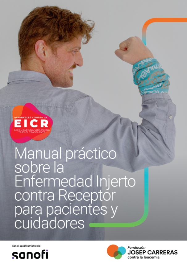
  • La Fundación Josep Carreras, en colaboración con SANOFI, también ha elaborado el primer Manual práctico sobre la Enfermedad Injerto contra Receptor para pacientes y cuidadores, así como material audiovisual para dar más voz a esta complicación. Todo este material estará disponible próximamente en la entrada del blog de la Fundación.

Hoy sábado 17 de febrero se celebra por primera vez el Día Mundial de la EICR, la Enfermedad Injerto contra Receptor. La EICR es una complicación que puede ocurrir tras un trasplante alogénico de progenitores hematopoyéticos (procedente de un donante), lo que se conoce popularmente como un “trasplante de médula ósea de un donante”. Este tratamiento constituye la única opción curativa en muchos casos de cánceres de la sangre, como la leucemia. La EICR sucede a entre el 30 y el 70% de los pacientes y, a pesar de los avances científicos, sigue siendo la principal causa de morbilidad y mortalidad en los pacientes trasplantados.

Por este motivo, la Fundación Josep Carreras se suma a la divulgación y conocimiento de esta complicación a través de diferentes acciones desarrolladas con la colaboración de SANOFI.

El pasado martes día 13, la Dra. Parody, miembro de la dirección médica de la Fundación Josep Carreras y directora médica del Registro de Donantes de Médula Ósea (REDMO), participó en la primera jornada organizada por la Asociación de pacientes de Linfoma, Mieloma, Leucemia y Síndromes Mieloproliferativos (AEAL) con el apoyo de Sanofi, en el Congreso de los Diputados. 

Este evento tenía como objetivo principal poner de manifiesto los retos a los que se enfrentan los pacientes con Enfermedad de Injerto Contra Receptor Crónica, la principal complicación tras un trasplante de médula ósea.
La Dra. Rocío Parody, en representación de la Fundación Josep Carreras y la Dra. Dolores Hernández, en representación de la Organización Nacional de Trasplantes (ONT) compartieron las principales conclusiones de la Cumbre Internacional en Trasplante organizada por la ONT el pasado mes de noviembre. Ambas pusieron de manifiesto el camino a seguir para el avance en la donación y el trasplante de órganos, tejidos y células a nivel global para los próximos diez años, así como sus implicaciones en el trasplante de médula.

*Si quieres ver la grabación de esta jornada clica AQUÍ

La EICR

La EICR es una complicación por la cual, tras el trasplante, las células trasplantadas reconocen al receptor (al paciente) como extraño y le atacan de diferentes formas e intensidad. En el trasplante a partir de un donante de progenitores hematopoyéticos, las células del donante constituirán el nuevo sistema inmune del paciente. Es por este motivo que, en este tipo de trasplante, serán las células del donante “las que puedan rechazar” a las células del receptor y, por ello, es esencial que donante y receptor tengan un genotipo idéntico.  Se trata de la combinación HLA, los antígenos leucocitarios humanos.

Cuando hablamos de “rechazo” tras un trasplante, en el imaginario colectivo está el trasplante de órganos sólidos, un proceso en el cual el sistema inmunitario del receptor de un trasplante ataca al órgano trasplantado. Sería el caso de un trasplante de hígado, de riñón, etc… En el caso de la médula ósea, es al revés. Es el “órgano trasplantado” en este caso, un tejido (la médula ósea) la que detecta que “ese cuerpo no es el suyo”. Por ello, la EICR se denomina así: Enfermedad del Injerto contra el Receptor.

La EICR aguda y crónica

La EICR puede ocurrir de forma aguda y rápida (habitualmente en los primeros 3 meses tras el trasplante) o crónica. No todos los pacientes tienen EICR aguda y/o crónica. Pueden desarrollar uno o los dos tipos o, ninguno. Hay que destacar que, a veces, no es tan fácil diferenciar la EICR aguda de la crónica ya que se “solapan”.

Los tres órganos clásicos” que se pueden ver afectados por la EICR aguda son, por orden de frecuencia: la piel, el tracto gastrointestinal y el hígado. A menudo, los pacientes presentan febrícula o fiebre precediendo al inicio de los síntomas de EICR aguda.

La EICR crónica puede aparecer como extensión de la EICH aguda, tras un intervalo libre de enfermedad o sin precedente agudo. Los síntomas de la EICR crónica pueden limitarse a un solo órgano o estar diseminados. A pesar de ello, se considera una enfermedad multisistémica. Es por ello por lo que la implicación de otros especialistas (dermatólogo, oftalmólogo, neumólogo, digestólogo, ginecólogo, urólogo…) es muy importante.

De forma más habitual, en la EICR crónica los principales órganos afectados son la piel, la boca, los ojos, los pulmones, el hígado y las articulaciones. Por lo general, no son afecciones que pongan en riesgo la vida del paciente, pero impactan de forma muy importante en la calidad de vida del paciente.

La incidencia de la EICR crónica es muy variable (30-80%). Su incidencia en niños es menor y aumenta en función de la edad del paciente. Entre los factores de riesgo para tener EICR crónica destacan el uso de sangre periférica como fuente de progenitores, el tipo de donante y el grado de disparidad HLA, la edad del donante y el haber padecido una EICR aguda previa. Generalmente, la EICR crónica aparece entre 3 meses y 2 años después del trasplante, pero puede surgir antes o, como hemos dicho anteriormente, “solaparse” con una EICR aguda.

Los principales factores de riesgo para desarrollar EICR crónica son:

  • haber desarrollado anteriormente EICR aguda.
  • la disparidad del HLA entre donante y receptor*
  • la edad del paciente
  • que la donación haya sido mediante sangre periférica

Los retos de la investigación

Habitualmente muchas de las áreas de investigación en hemopatías malignas están ligadas a entender los procesos por los cuales se desarrollan estas enfermedades: leucemias, linfomas, mieloma múltiple… Pero es fundamental enfocar líneas de investigación también a preservar la calidad de vida tras un tratamiento como un trasplante de médula ósea.

En el abordaje actual de esta patología, las opciones de tratamiento siguen siendo limitadas. Así, el tratamiento de primera línea consiste en inmunosupresores y corticosteroides, con una tasa de respuesta de entre el 50% y el 60%. Aproximadamente la mitad de los pacientes que reciben corticoides no responderán al tratamiento o recaerán al intentar retirar la medicación, y necesitarán una segunda línea de tratamiento. Cabe decir que estos pacientes tienen una inmunosupresión importante y, además, pueden tener otras complicaciones graves además de la EICR crónica.

Debido a la frecuencia e impacto de la EICR en el paciente trasplantado, y que tras el tratamiento de primera línea ni hay un consenso uniforme ni todos los pacientes responden adecuadamente, esta enfermedad tiene un amplio campo de investigación tanto para prevenirla como para tratarla. Igualmente hay estudios dirigidos a los factores de riesgo más específicos que los ya conocidos, como pueden ser marcadores en sangre que sean predictores para el paciente de desarrollar la enfermedad, pero estos últimos no están aún tan avanzados y sobre todo en la práctica clínica no son fáciles de instaurar. 

* 3 de cada 4 pacientes que necesitan un trasplante de médula ósea compatible, no disponen de un donante entre sus familiares. En esos casos, en España, es el Registro de Donantes de Médula Ósea (REDMO), programa de la Fundación Josep Carreras quien, en coordinación con la Organización Nacional de Trasplantes y las Comunidades Autónomas, busca un donante HLA compatible alrededor del mundo. Entre los más de 40 millones de donantes de médula ósea disponibles en los registros internacionales. Para tener una “compatibilidad idéntica”, el donante y el receptor deberán haber heredado el mismo conjunto de antígenos (el sistema HLA) de cada uno de sus padres.

Tal como comentábamos en titulares, desde la Fundación Josep Carreras hemos creado el primer Manual práctico sobre la Enfermedad Injerto contra Receptor para pacientes y cuidadores que incluye todo lo relativo a la EICR (¿qué es?, ¿por qué ocurre?…), cuáles son sus manifestaciones agudas y crónicas así como trucos para sobrellevar sus efectos.

¡GRACIAS A SANOFI POR SER IMPARABLES CONTRA LA EICR!

Primera conferencia de alto nivel sobre la discriminación financiera de los supervivientes de cáncer en Bruselas

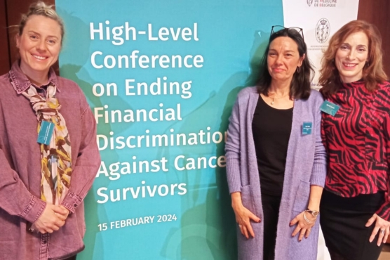
Hoy, 15 de febrero de 2024, se celebra en Bruselas, la primera conferencia de alto nivel sobre el derecho al olvido oncológico que sufren muchos supervivientes de cáncer en materia de productos financieros y de seguros.

Este evento tiene como objetivo abordar los desafíos financieros a los que se enfrentan los supervivientes de cáncer, así como acentuar la importancia de disponer de un código de conducta común entre países de la Unión Europea. Entre los participantes, destacan los ministros de Sanidad y Economía de Bélgica; los comisarios europeos de Salud y Seguridad Alimentaria, y de Servicios financieros; miembros jurídicos del Tribunal Europeo de los Derechos Humanos; supervivientes de cáncer; responsables políticos nacionales y de la Unión Europea, y representantes de compañías de seguros. Todos ellos discutirán los obstáculos para obtener seguros o préstamos y compartirán las mejores prácticas para eliminar la discriminación contra los sobrevivientes de cáncer en la Unión Europea.

La Fundación Josep Carreras participa activamente en esta jornada con la contribución de Elordi García Insausti, superviviente de una leucemia aguda y Alexandra Carpentier, responsable del Programa de Experiencia del Paciente de la organización y miembro de la comisión ejecutiva de la Federación Catalana de Entidades contra el Cáncer (FECEC). Esta última planteará los retos que está suponiendo la aplicación del Real Decreto Ley para regular el Derecho al Olvido Oncológico que se aprobó en España el pasado 29 de junio de 2023.

Este evento europeo ha sido organizado por la asociación Ending Discrimination Against Cancer Survivors, con el apoyo de Fonds Cancer, la Real Academia de Medicina de Bélgica, y bajo los auspicios de la Presidencia belga del Consejo de la Unión Europea.

Más información en: https://endingdiscrimination-cancersurvivors.eu/high-level-conference-on-ending-financial-discrimination-against-cancer-survivors/

A new immunotherapy against multiple myeloma shows more effectiveness in experimental models than CAR-T already in use

Recent research by Dr. Pablo Menéndez’s lab at the Josep Carreras Institute, the Spanish National Cancer Research Center (CNIO) and the University Hospital October 12 shows how a new immunotherapy against multiple myeloma based on STAb cells is more effective in experimental models than the current CAR-T.

Immunotherapy is already improving treatment options for many cancers, but research groups continue to explore ways to boost the body’s immune system and use it against the tumour.

Researchers at the Josep Carreras Leukaemia Research Institute, the Spanish National Cancer Research Centre (CNIO) and the University Hospital 12 de Octubre have developed a new immunotherapy for multiple myeloma that has proved to be more effective in the laboratory than the current preferred immunotherapy.

The new immunotherapy is based on so-called STAb cells. It has been tested only in experimental models, so it has yet to pass clinical trials in humans and will therefore take at least two years before it reaches the clinic.

The study, published in the journal Science Translational Medicine involved Dr Clara Bueno and Dr Aida Falgas, from the Stem Cell Biology, Developmental Leukaemia and Immunotherapy group, and was coordinated by Dr. Luis Álvarez-Vallina head of the H12O-CNIO Cancer Immunotherapy Clinical Research Unit. It is a collaboration between this unit and the Josep Carreras Institute; the Hospital Clínic in Barcelona; and the Universities of Salamanca and Complutense of Madrid.

STAb cells
Multiple myeloma is the second most common haematological cancer in adults, after lymphomas. In 2020, 176,404 new cases of multiple myeloma were diagnosed worldwide and its incidence is estimated at 1.78 per 100,000 people.

In recent years, these cancers are beginning to be treated with CAR-T cell immunotherapy,” explains Luis Álvarez-Vallina, “which has been a substantial improvement over the therapeutic tools that existed before. Despite this, and although patients now have longer survival times, it is a disease in which a significant proportion of patients relapse, so relapse treatments are necessary.

Advantages over conventional CAR-T
CAR-T cell therapy, whose full name is chimeric antigen receptor T-cell therapy , involves modifying a patient’s T-cell immune cells (white blood cells) in the laboratory so that they are able to recognise and fight tumour cells.

Research has compared this treatment with another cellular immunotherapy based on or STAb-T cells, which can be considered an evolution of CAR-T cell therapies. In both cases, the laboratory-modified cells recognise the same antigen, called BCMA, which is only present on tumour cells. In this way, the modified cells target and attack only cancer cells.

The study now presented in Science Translational Medicine shows that STAb-T cells outperform CAR-T cells, because they can make other T cells in the body, which have not been modified, also fight cancer cells, thus amplifying the effect of the therapy.

In addition, STAb-Ts overcome an element that slows down CAR-Ts. In some patients with multiple myeloma, the BCMA antigen – which identifies tumour cells – is found in soluble form when there is a high tumour burden. It turns out that the fact that the antigen is soluble prevents the activity of CAR-T cells, but does not affect STAb-T cells, the now-published research shows.

Immune memory
“Finally, we also showed that STAb-T cells generate immunological memory”, says Álvarez-Vallina. After recreating the disease in model animals and treating them with STAb-T cells, the team obtained cells from various organs, mainly spleen and bone marrow, and observed the generation of memory STAb-T cells.

“This is very important”, explains Álvarez-Vallina, “because we know that the persistence of CAR-T cells in the body, i.e. immunological memory, is related to the extent of the anti-tumour effect and, therefore, to better control of the disease”.

The research group aims to carry out a clinical trial in collaboration with the 12 de Octubre University Hospital to treat people with multiple myeloma with this new STAb-T immunotherapy.

Researchers at the Josep Carreras Institute test an affordable and reliable genetic screening for leukaemia patients

Researchers from the Myelodysplastic Syndromes (MDS)’s group at the Josep Carreras Leukaemia Research Institute have successfully tested a new method to determine cancer-related genetic variants in pooled samples from patients. This new method improves the current method, based on targeted next generation sequencing technology, to deliver a cost-effective diagnostic of cancer patients.

Over 90% of patients diagnosed with MDS bear genetic variants in their DNA which confers them special susceptibility towards cancer or are cancer-promoting themselves. Thus, in the last decades, the international researcher’s community has been deciphering the most relevant genetic variants for each cancer type and how can this knowledge be used to better manage these patients more effectively.

In this sense, molecular insight is used in the clinical practice not only in cancer diagnostic but also in treatment guidelines. In MDS, the preferred methodology to interrogate genetic alterations is multiplexed targeted Next Generation Sequencing (tNGS). However, tNGS is expensive and technically complex and, therefore, cannot yet become an international standard when it comes to analysing lots of samples.

To address this issue, a team of researchers from the Josep Carreras Leukaemia Research Institute has been testing an alternative methodology combining tNGS and classical Sanger DNA sequencing, a much cheaper technology, in MDS patients. The team, spearheaded by Dr. Oriol Calvete and supervised by Dr. Francesc Solé, hypothesized that pooling samples from different MDS patients would still allow for the precise detection of characteristic mutations using tNGS, despite the dilution effect.

In a recent publication at the scientific journal Biomedicines, the team proved that pooling samples from MDS patients had little impact on the sensitivity of tNGS and that up to 4 samples could be successfully processed in a single experiment, without losing any quality in the outputs. After the identification of any concerning genetic variant in the pool, the precise assessment of the patient bearing it can be easily resolved by direct sequencing using the Sanger method, an old and reliable technology more than twenty times cheaper than tNGS.

This pilot experience in MDS patients demonstrates that the affordable screening for cancer-related genetic variants is possible over a large number of samples, while keeping the accuracy of tNGS. In this way, the door opens to the widespread adoption of precise and reliable genetic screening of patients, internationally, allowing for a better patient stratification and helping clinicians choose the best treatment option.

Discovered how to predict whether patients with leukaemia will be sensitive to epigenetic drugs

Researchers from the Josep Carreras Leukaemia Research Institute, spearheaded by the Cancer Epigenetics group led by Dr. Manel Esteller, described the effects of chemotherapy at the single cell resolution in the bone marrow of Myelodysplastic Syndrome patients. The results help better understand which cellular populations are more sensitive to azacitidine, an epigenetic drug, and offer valuable insights on how to treat relapses after treatment.

Alterations in the chemical modifications that control gene expression, known as Epigenetics, have proven to be one of the most characteristic properties of all human tumours. This realization has led to the development of very intense pharmacological research to find drugs that act at this level against cancer. Today, there are nine epigenetic drugs approved for use in oncology, especially in leukaemia, lymphomas and soft tissue tumours. However, a mystery remains: Why do some of these patients respond clinically to these compounds while in other cases they show resistance to their action?

A recent article authored by Dr. Ignacio Campillo and Marta Casado, researchers from the Cancer Epigenetics led by Dr. Manel Esteller, Director of the Josep Carreras Leukaemia Research Institute, ICREA Research Professor and Chairman of Genetics at the School of Medicine of the University of Barcelona, provides the first answer to this question by showing how the persistence of certain cancer cells, bearing specific mutations, is associated with the lack of clinical benefit of these pharmacological principles.

The research, published at the journal Cancer Research Communications, the official journal of the American Association for Cancer Research (AACR) for the rapid publication of interesting results, is a joint effort of several groups at the Josep Carreras Institute, including single cell specialists Dr. Elisabetta Mereu and Dr. Caterina Mataand clinical researchers Dr. Lurdes Zamora, Dr. Blanca Xicoy, Dr. Gaël Roué and Dr. Franscesc Solé.

“We decided to focus our study on a type of bone marrow cancer, where leukaemia blood cells are formed, called myelodysplastic syndrome because the treatment of choice is an epigenetic drug called azacitidine, an inhibitor of DNA methylation. We studied what was happening at the DNA and protein levels in thousands of cells separated from these patients at two time points: before and after receiving the epigenetic therapy”, explains Dr. Esteller about the research.

“We managed to characterize more than 30 cell subtypes and 50 genes, observing that the patients where pharmacological treatment had an effect had a particular profile at the individual cell level: they presented a decrease in the number of mutations in stem and progenitor cells and in immature granulocytes and monocytes. This suggests that if we do not eliminate these altered primitive cells, which appear early during the tumour process, the therapy has little chance of success.

The good news for patients resistant to epigenetic drugs is that we have detected that some of the new mutations that appear may now be targets of other drugs specifically directed against them. As if it were a game of cat and mouse between the doctor and the cancer, the tumour’s strength against one drug generates its vulnerability against another drug.

Hence the importance of molecular studies at the single cell level that allow us to predict not only the prognosis of the disease but also what this leukaemia may be sensitive to”, concludes the researcher.

The research has received funding from the Spanish Ministry of Science and Innovation, the Marie Skłodowska-Curie Action, the CELLEX Foundation and “La Caixa” Foundation among others.