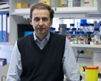
Anna Bigas, new Deputy Director of Preclinical Research at the Josep Carreras Leukemia Research Institute

Dr. Bigas, well-recognized international researcher in the study of the generation and renewal of stem cells and their role in cancer processes, assumes the functions of Deputy Director of Preclinical Research at the Josep Carreras Leukemia Research Institute.

Dr. Bigas has joined as Deputy Director of Preclinical Research at the Josep Carreras Institute, maintaining her relationship with the Hospital del Mar Medical Research Institute (IMIM), where she leads the Stem Cells and Cancer group. The new responsibility that Dr. Bigas assumes will serve to enhance coordination between all groups in the institute that do basic and translational research, seeking synergies between them and optimizing their research, as well as to enhance the existing collaboration between the professionals of our institute and those of the IMIM.

 In the last ten years, as head of the Stem Cells and Cancer group, she has studied the molecular and physiological processes of stem cells in different tissues, in order to draw similarities with the appearance and progression of cancerous processes.


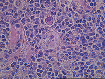
In particular, her research focuses on the study of hematopoietic stem cells, responsible for the formation of blood cells, in order to understand the process of leukemia and develop strategies to fight against this disease. Currently, her research group is working on the identification of epigenetic and gene expression profiles, which allow discovering new therapeutic targets not yet exploited clinically. 

Research trajectory 

Dr. Bigas holds a PhD in cell biology from the University of Barcelona, ​​where she began her research in the generation and renewal of stem cells. During the 1990s, she developed her career as postdoctoral researcher at the Fred Hutchinson Cancer Research Center (Seattle, USA) where she identified the role of the Notch signaling pathway during hematopoietic differentiation. Subsequently, she created her own research group at the Oncology Research Institute (IRO / IDIBELL), where she intensified the study of the Notch, Wnt and NFkB signaling pathways and their molecular mechanisms, involved in the regulation of hematopoietic system and its oncogenic transformation (1997-2008). Since 2009 she has been leading the Stem Cells and Cancer group at the Hospital del Mar Medical Research Institute (IMIM).


Currently, Dr. Bigas is the Scientific Director of CIBERONC (Institute of Health Carlos III) where she coordinates the collaborative research activities in cancer of 50 research groups, distributed throughout the state. She is also a member of the management of the International Society of Experimental Hematology (ISEH) and member the Spanish Association of Hematology and Hemotherapy (AEHH), the European Hematology Association (EHA) and the Spanish Association for Cancer Research (ASEICA). Due to her international relevance, she is reviewer for the European Research Council (ERC) and the Research Councils of Finland and Lithuania. She also occasionally serves as reviewer for the Norwegian Research Council, the Medical Research Council of the United Kingdom, the French National Research Agency and the Israeli Ministry of Science.


New epigenetic targets identified for the potential treatment of multiple myeloma

Researchers at the Josep Carreras Leukaemia Research Institute have identified epigenetic alterations in multiple myeloma and have tested a compound that reverse those alterations, opening the door to a new therapeutic drug in the treatment of multiple myeloma.

Multiple myeloma is an incurable type of cancer representing 1% of all cancers. It occurs when a type of blood cell (plasma cells) expand uncontrollably in the bone marrow. Most myeloma patients suffer from skeletal-related complications during the course of the disease, such as severe bone pain, pathological fractures and others, that affect both the quality of life and their overall survival. Mesenchymal stromal cells are essential in the formation and function of the bone marrow and the bone itself. It is well-documented that mesenchymal stromal cells from multiple myeloma are altered and contribute to the progression of the disease.

Dr Esteban Ballestar’s group has just published a study in the journal Nature Communications reporting the identification and characterization of epigenetic alterations in mesenchymal stromal cells from multiple myeloma patients that are associated with the altered functions of these cells in myeloma bone disease.

Such alterations do not modify the cell’s genetic information but their capacity to use it, causing an abnormal behaviour in the affected cell.

The team has demonstrated that cancerous plasma cells are able to induce those epigenetic alterations in mesenchymal stromal cells that result in their diminished capacity for bone formation.

In addition, the team has identified a compound, named CM-272, previously developed by Dr Felipe Prósper’s team, that can reverse those epigenetic alterations and the altered functions of mesenchymal stromal cells from myeloma patients. The identification of these alterations, and the use of CM-272 to reverse them, may represent an important advance towards new therapeutic options in the treatment of multiple myeloma.

This study, whose first authors are Dr. Antonio Garcia-Gomez and Dr. Tianlu Li, was completed in collaboration with the teams of Dr. Mercedes Garayoa (Cancer Research Center, Salamanca), Dr. Felipe Prosper and Dr Xabier Agirre (Center for Applied Medical Research, Navarra) and Dr Manel Esteller (Josep Carreras Leukaemia Research Institute).

This research has been funded by the Asociación Española contra el Cáncer (AECC), that funded Dr Antonio Garcia, the Multiple Myeloma Research Foundation and the Spanish Ministry of Science and Innovation, co-financed by FEDER funds.

Discovered the explanation of the patients with “lazarus-like” cancer

An article published in Cancer Cell with the collaboration of the group of Dr. Manel Esteller, Director of the Josep Carreras Leukaemia Research Institute, ICREA Research Professor and Professor of Genetics at the University of Barcelona, ​​today provides us with the reasons that would explain the extraordinary response of these individuals to cancer therapy.

There is a very special subgroup of cancer patients. These are people who have advanced, and highly aggressive cancer mostly associated with a very short survival time, but surprisingly they respond very well to treatment, greatly increasing their life expectancy. These patients are colloquially called “Lazarus” because, being on the verge of death, they return to full life, as happened with the person of the same name described in the biblical texts. These “Lazarus” patients represent a minimal proportion of individuals with cancer and until now the causes of this phenomenon were unknown.

“The study has been led by the National Cancer Institute (NCI) of the United States, which are world pioneers in genomic studies of tumours. They had managed to gather more than 100 exceptional responding patients, where they were obtaining the mutations of the tumours, and they asked us to also analyse their epigenome and we did so. It has been a work of several years and intense, but the discoveries obtained have been worth all the effort: we have obtained the first explanation of why these patients evolve so well. ” – states Dr Manel Esteller, co-author of the Cancer Cell study – “It turns out that the cancers of these people have randomly accumulated several genetic and epigenetic defects that make them hypersensitive to the drug that the doctor gives them. It would be as if on a floor that has just been polished, a banana skin will also be deposited by chance: the fall of a person walking by would occur for sure. Well, that is what happens to these tumours: they accumulate two or three events that derail the growth of the cancer when it receives the drug. An example: a high-grade brain tumour has a genetic lesion in a DNA repair gene and a mutation in a different gene that also repairs DNA. That patient, if he receives a drug that damages DNA, won’t be able to fix that defect and that tumour won’t be able to grow.” -explains Dr Esteller and adds- “Now the dream is that this knowledge serves to help other patients with tumours of much more common clinical behaviour. For example, we could think that the external induction of a second alteration in a pathway already damaged in the tumour, would create a “Lazarus” patient that would respond very well to a drug designed against that cellular pathway. “

New advances in acute lymphoblastic leukaemia in adults

Acute lymphoblastic leukaemia (also known as acute lymphocytic leukaemia or ALL) is a type of cancer of the blood in which, for unknown reasons, excessive amounts of immature lymphocytes (lymphoblasts) are produced. The cancerous cells multiply rapidly and displace the normal cells of the bone marrow, the soft tissue inside some bones where blood cells are formed.

Although ALL often mainly affects children, it is not uncommon to observe it in adolescents and young adults. In adults, this kind of leukaemia prevails in young males (average age between 25 and 30 years old). Only 10-15% of patients are over 50 years old. In Spain, the annual incidence of ALL in adults is 30 new cases per million inhabitants per year.

Dr Ribera’s group has led adult ALL treatment throughout Spain for 20 years.

Recently they have published two very important works on aspects of this disease:

-TRANSPLANT: On one hand, a study published in Blood, one of the most important scientific journals on haematology in the world. Entitled Do all adults with acute lymphoblastic leukaemia have to be transplanted? The answer is: no.

In this work, they have been able to identify which patients will need a transplant of haematopoietic stem cells for their recovery and which ones do not. To do this, they have studied 348 adult patients with acute lymphoblastic leukaemia and have seen that 218 will not need to have a transplant (and will continue with chemotherapy) and 106 who will.

The assessment is based on the detection of the minimal residual disease, which is what the patient has “hidden” in the body and the researchers cannot detect with a microscope, but they can with other systems. If the quantity of these cells is small, a transplant would not be necessary, but if the quantity is high, it will be. 

In this study they have worked together with Dr Alberto Orfao, member of the Scientific Committee of the Josep Carreras Institute and Director of the General Cytometry Service of the University of Salamanca and expert in the detection of residual disease. Dr Orfao’s team has very sensitive technology, called next generation flow cytometry, which enables these cells to be detected to quantify them very precisely, which has allowed for improvement in clinical decisions depending on the figures obtained with this cutting-edge technology.

-RELAPSES IN T-CELL ACUTE LYMPHOBLASTIC LEUKAEMIA IN ADULTS: On the other hand, Dr Eulalia Genescà and Dr Josep Maria Ribera have worked together on the study, published in the Genome Biology journal, with the laboratory of Núria López-Bigas at IRB Barcelona and with Anna Bigas’ groups at the Hospital del Mar Medical Research Institute.

It is a genetic study of a type of acute lymphoblastic leukaemia, the T-cell type, which despite initially responding to treatment positively, has frequent relapses and a bad prognosis.

The group has studied how these patients were genetically upon diagnosis and at the time of relapse. The team has discovered that the cells responsible for resistance to treatment of T-ALL leukaemia in adults are already present in the tumours before the first diagnosis.

To carry out this work, they have sequenced the genome of samples from 19 patients at the time of diagnosis as well as during treatment and upon relapse, and they have compared the information obtained with a database of 238 patients (adults and children) affected by similar tumours. Through the comparison of the primary leukaemias of the adults and those of the paediatric patients, they could observe that the mutational processes are similar, although the specific genetic alterations responsible for the relapse are different in each individual case. For this reason, an algorithm is being developed to classify these relapses, as not all of them are the same. Sometimes the patient can relapse from the same disease they initially had, and other times it is seen that upon relapse the leukaemia has changed.

This has permitted in-depth study of the matter of relapses and their complexity. Almost every relapse is different, which has major implications for designing the treatment. For this reason, the team will continue to be UNSTOPPABLE to continue getting to know them in more detail.

Researchers from the Josep Carreras Institute received one of the five grants awarded worldwide by the Facebook founder’s Foundation to study COVID-19

In May, the epigenetics and immune diseases group of the Josep Carreras Leukaemia Research Institute, led by Dr Esteban Ballestar, together with Roser Vento-Tormo (Sanger Institute, United Kingdom), obtained one of the five worldwide grants awarded by the Chan Zuckerberg Initiative, a non-profit organisation owned by the Facebook founder, Mark Zuckerberg, and his wife, Dr Priscilla Chan. Each of the five groups that received this grant has worked independently on a specific theme relating to COVID-19.

The general objective of all the groups was to make an atlas of patients with COVID-19 studying different types of tissues (blood, nasal biopsies, pharynx biopsies, etc) to obtain an atlas of all the alterations of the gene expression that these patients have with different subthemes.

The five groups have recently made the first draft of the results available to the whole scientific and medical community worldwide.

In record time, Dr Ballestar’s group has generated the atlas belonging to COVID-19 patients who had a pre-existing immune disease (such as immunodeficiencies, rheumatoid arthritis, etc) to see whether these individuals have a more deficient or altered response compared with patients who have had COVID-19 but who do not have any pre-existing medical conditions.

The group is currently analysing these data and preparing a report with the conclusions of this study.

Each cell is different because, although they have the same genome, each one expresses a series of genes to be a specific cell type (a B lymphocyte, a macrophage, etc.). In response to different situations, what these cells are expressing also changes a little.

To do this work, the group has used single-cell technology to obtain the map of expressions of thousands of immune cells of each patient to understand how they communicate and cooperate with each other in response to the SARS-CoV-2 infection and to understand how these genes that each cell expresses before the presence of the SARS-CoV-2 virus are different genes between those individuals who have altered immunity and those who are healthy.

The Josep Carreras Institute is the only one in Spain that can use this technology for this kind of study because the equipment is in a special biosecurity chamber, which is what enables it to do so.

A new study from Josep Carreras Institute reveals a new prognostic marker in a specific subtype of acute lymphoblastic leukemia (ALL) with adverse outcome in infants

Acute lymphoblastic leukemia (ALL) is the most common type of children cancer and, in some cases, presents with a very poor prognosis in infants younger than one year. Despite the increase of survival chances in pediatric patients with ALL during the last decades, deeper studies of the mechanisms involved in this disease are required, especially in a subgroup of patients with an aberrant genetic alteration (known as t(4;11)).

The “Lymphocyte Development and Disease” group, from Josep Carreras Leukaemia Research Institute, led by Dr. Maribel Parra, has recently published a new paper in Leukemia journal, describing HDAC7 protein as a prognostic marker of this specific subtype of infant ALL. In normal conditions, HDAC7 drives the appropriate formation of B lymphocytes, key cells of the immune system. Moreover, it prevents B lymphocytes precursor cells from acquiring an uncontrolled proliferation, possibly conducting to leukemia onset. The study published demonstrates that HDAC7 expression is almost absent in infants diagnosed of ALL with t(4;11) alteration. Instead, other subgroups with a more favorable outcome, display higher levels of HDAC7.

“We have observed that, when we introduce HDAC7 protein in cells derived from these patients, leukemic cells stop proliferating in an aberrant manner and, most of them, undergo cell death processes. Moreover, data indicate a direct relationship between lack of HDAC7 and chemotherapy resistance, which is very common in these infant patients who, in case of surviving, usually present recurrences”, explains Oriol de Barrios, postdoctoral researcher of the group and first author of the study. “Given these results, which set HDAC7 as a novel marker of good prognosis in t(4;11) infant ALL subtype, our next goal is to find new therapeutic strategies that conduct to a precisely directed HDAC7 induction. We believe that HDAC7 is a promising therapeutic target and that an adequate pharmacologic modulation may considerably increase survival chances of this aggressive subtype of infant ALL”, states Dr. Maribel Parra, who has coordinated the study.

The study has been conducted with an extensive network of collaborators around Europe, from countries like Greece, Italy and the Netherlands, as well as with the participation of Dr. Pablo Menéndez research group, also from the Josep Carreras Institute.

Dr. Marcus Buschbeck leads the European Network INTERCEPT-MDS focusing on the shared training and research in Myeloid Diseases

►Dr. Marcus Buschbeck, group leader of the Josep Carreras Leukaemia Research Institute, coordinates the Innovative Training Network (ITN) INTERCEPT-MDS, a Marie Skłodowska Curie Action, funded by the European Commission under H2020 framework programme.


INTERCEPT-MDS intertwines 17 public and private centers from 7 countries to train 12 early-stage researchers (ESRs), at the PhD student level, with the aim to deepen on disease interception, a novel concept referring to the treatment of a disease before it fully develops by removing altered cells.

To make disease cell interception a reality, researchers need to overcome two key challenges: First, to identify few altered disease cells among many healthy ones. Second, the development of strategies that allow specific targeting of malignant cells without affecting healthy cells.

In the INTERCEPT-MDS ITN, Marcus Buschbeck and his colleagues propose to approach these challenges through shared research and multidisciplinary and multisectoral training of 12 ESRs. The project is aligned with overall goals of the EU LifeTime Initiative of which the Josep Carreras Leukaemia Research Institute is an associated partner.

With the aim to train and present Europe’s first experts in the new field of disease cell interception, the ITN will take advantage of single-cell omics methods that have reached a level of maturity to be applied to a broad-scale.

For interception, researchers will explore and exploit the epigenetic regulatory space by leveraging privileged access to tool compounds and by performing genetic loss-of-function screenings in vivo and in vitro. This project will focus on myeloid diseases because they are a suitable paradigm for clonally evolving conditions and come with a significant advantage. In contrast to most other diseases is the availability of stem cells, niche cells, and their progeny through samples of the clinical routine.

The Josep Carreras Leukaemia Research Institute will coordinate the network that includes as ESR-recruiting beneficiaries the Chemotherapeutisches Forschungsinstitut Georg-Speyer-Haus Stiftung and the Technischen Universität München from Germany, the Veterinärmedizinische Universität Wien from Austria, the Institut National de la Santé et la Recherche Médicale from France, the Università Degli Studi di Firenze from Italy, the Erasmus Universitair Medisch Centrum Rotterdam from The Netherlands, the Universitetet i Bergen from Norway as well as the companies Biobam Bioinformatics from Spain and the Müncher Leukämielabor GmbH from Germany.

Additional partners contributing to the research and training programme are the Universitat Autònoma de Barcelona, the management trainers from Leadership Sculptor GmbH, the biotech company Aelian Biotechnology from Austria, the patient support association Connaître et Combattre les Myélodysplasies from France, the Université de Paris, the University of Florida, the Universitätsklinikum Aachen from Germany and the genomic service provider GenomeScan B.V. from The Netherlands.

*This project has received funding from the European Union’s Horizon 2020 research and innovation programme under the Marie Skłodowska-Curie grant agreement No 953407

El Instituto Josep Carreras participa en el Congreso Anual de Hematología en España

La semana del 26 al 30 de octubre se celebró la LXII edición del congreso de la Sociedad Española de Hematología y Hemoterapia y la XXXVI del congreso de la Sociedad Española de Trombosis y Hemostasia.

Debido a la situación de la pandemia de COVID-19, esta edición se tuvo que llevar a cabo de forma telemática. Aun así, el congreso fue todo un éxito alcanzando la cifra de 2.300 asistentes.

Este congreso se celebra de forma anual y en él se desarrollan diferentes actividades, destacando:

Los simposios oficiales, que tienen como objetivo difundir los últimos avances en investigación clínica y traslacional entre los profesionales de la hematología. Para ello se invita a expertos nacionales e internacionales en cada tema.

Las comunicaciones de trabajos científicos, en las que los profesionales dedicados a a Hematología presentan las novedades de sus trabajos de investigación básica, traslacional o clínica. Existe un comité de evaluación de estas comunicaciones para clasificarlas, según su calidad, en comunicaciones orales (cuando se consideran más relevantes) o en comunicaciones que se exhiben como pósters informativos para su lectura.

El Instituto Josep Carreras estuvo presente de forma muy notable a través de diferentes trabajos presentados.

Complicaciones en el trasplante de progenitores hematopoyéticos

El doctor Enric Carreras, director del Registro de Donantes de Médula Ósea gestionado por la Fundación Josep Carreras e investigador del grupo Barcelona Endothelium Team (BET) del Instituto Josep Carreras centra su investigación en las células que recubren la cara interna de los vasos sanguíneos (las venas, las arterias y los capilares) que se llaman células endoteliales y forman el mayor órgano del cuerpo humano: el endotelio.

En el simposio del congreso, el Dr. Carreras expuso el problema de la microangiopatía trombótica en el trasplante de progenitores hematopoyéticos. Existe un grupo de enfermedades cuyos síntomas tienen un origen común: la lesión en el tejido endotelial que recubre el interior de los vasos sanguíneos. Entre otras destaca la llamada MAT (Microangiopatía Trombótica). Es una complicación infrecuente pero muchas veces mortal del trasplante que provoca una progresiva oclusión de los pequeños vasos sanguíneos (capilares) por plaquetas y otros componentes de la coagulación. Esta oclusión dificulta el paso del resto de componentes sanguíneos, como los glóbulos rojos que se rompen al pasar aumentando progresivamente la oclusión que puede llegar a ser casi completa. Este suceso provoca una serie de síntomas como anemia, trombopenia, hipertensión y fallo en órganos como el riñón, el sistema nervioso central, el cardiovascular o el gastrointestinal que pueden llegar a ser letales. Por ello, el BET centra parte de su investigación en la detección precoz de esta complicación tras el trasplante de progenitores hematopoyéticos y la administración de tratamientos que puedan evitar la elevada mortalidad asociada a esta patología.

Nuevas tecnologías aplicadas al diagnóstico y tratamiento del linfoma

El doctor Tomás Navarro, investigador principal del grupo de neoplasias linfoides del Instituto Josep Carreras coordinó, junto al Dr. Antonio Gutiérrez del Hospital Son Espases de Mallorca, un simposio acerca de las nuevas tecnologías aplicadas al diagnóstico y tratamiento del linfoma difuso de célula grande B.

En este simposio se invitó al Dr. Georg Lenz, jefe del servicio de Hematología del Hospital Universitario de Münster (Alemania) que habló de la importancia de la investigación traslacional con el objetivo de encontrar tratamientos dirigidos contra dianas terapéuticas.

También se abordó el tema de la biopsia líquida que trata de detectar material genético de las células del linfoma en la sangre circulante. Las células tumorales vierten en la sangre tanto ADN como ARN, y por tanto analizando este material genético podemos tener una representación del tumor en la sangre. Aunque seguramente no sustituirá a la biopsia de tejido, sí que permite estudiar a los pacientes de una manera más completa, teniendo un mapa más concreto y detallado de la enfermedad.

En este simposio también se mostraron los nuevos tratamientos existentes para este tipo de linfoma difuso de célula grande B, muchos de los cuales de momento se utilizan para pacientes que no responden a las primeras líneas de tratamiento, como son:

Los anticuerpos monoclonales. Cuando hablamos de anticuerpo nos referimos a una proteína del organismo que se adhiere a otra proteína específica llamada antígeno. Los anticuerpos circulan por todo el cuerpo hasta que encuentran y se adhieren al antígeno. Una vez unidos, pueden forzar a que otras partes del sistema inmunitario destruyan a las células que contienen ese antígeno. Hoy en día se pueden diseñar anticuerpos que tengan como objetivo específico un antígeno concreto.

Los anticuerpos conjugados, que se elaboran con un anticuerpo monoclonal químicamente ligado a un medicamento.

Los anticuerpos biespecíficos, que por un lado se unen a la célula tumoral y por otro lado estimulan células para atacar al tumor.

Los CAR-T, un tratamiento que consiste en extraer las células T del paciente (las encargadas de la defensa de nuestro organismo), modificarlas en laboratorio y volverlas a infundir al paciente. Esta modificación permite que estas células ataquen los receptores que se encuentran en la membrana de las células tumorales (llamados antígenos “CD”) y las eliminen.

Premio al Mejor Caso

El Club Español de Citología Hematológica organiza el simposio Casos clínicos citológicos, uno de los de mayor éxito de asistencia (este año alcanzando la cifra récord de más de 700 asistentes), expone 6 casos interesantes desde el punto de vista diagnóstico y/o terapéuticos que pueden tener un especial interés. Este año se presentaron inicialmente24 casos y el comité de selección del simposio escogió a 6 para que fueran presentados en el congreso. El ganador del Premio al Mejor Caso recayó en un trabajo colaborativo del Instituto Josep Carreras.

En este caso, presentado por la doctora Maria Huguet, han colaborado diferentes investigadores el Instituto Josep Carreras como las doctoras Blanca Xicoy y Lurdes Zamora del grupo de neoplasias mieloides, el Dr. Marc Sorigué del grupo de citómica funcional y el Dr. Navarro del grupo de neoplasias linfoides.

Trataba el caso de un paciente de la doctora Blanca Xicoy que fue diagnosticado de policitemia vera y al cabo de los años se vio que el paciente iba a peor con el tratamiento administrado. Por ello, se decidió parar el tratamiento y al cabo de 1 mes el paciente empeoró más, como si se estuviera transformando a una leucemia mielomonocítica crónica. El punto fuerte de este caso es que se estudió todo desde un punto de vista molecular, las mutaciones de la enfermedad en el diagnóstico y en el momento de la evolución de esta enfermedad en este paciente. También se separaron los monocitos mediante un citómetro y se demostró que estas células presentaban las mismas mutaciones que tenía la enfermedad Policitemia vera en el diagnóstico, pero que habían aumentado mucho.

Este caso ilustra la utilidad de las nuevas tecnologías NGS (Next Generation Secuency) que pueden llegar a tener un papel crucial en el diagnóstico y el seguimiento de estas enfermedades.

Las comunicaciones orales

Por lo que respecta a comunicaciones orales, el Dr. Josep Maria Ribera, investigador principal del grupo de leucemia linfoblástica aguda, presentó los resultados preliminares de un ensayo clínico llamado Ponalfil del grupo PETHEMA en el que combinaron el fármaco Ponatinib y quimioterapia en adultos jóvenes con LLA con cromosoma Filadelfia.

La combinación de quimioterapia e inhibidores de tirosincinasa como Ponatinib ha mejorado el pronóstico de estos pacientes y los resultados preliminares de este ensayo indican una alta eficacia antileucémica a corto plazo, con una toxicidad aceptable.

Este estudio, junto a otros en curso, contribuirá probablemente a indicar Ponatinib como tratamiento de primera línea en la LLA con cromosoma Filadelfia.

La Dra. Christelle Ferrà, del Servicio de Hematología Clínica del Instituto Catalán de Oncología, presentó la experiencia del Grupo Español de Trasplante Hematopoyético y Terapia Celular (GETH) acerca de la evolución de los pacientes con leucemia linfoblástica aguda que recaen tras un trasplante alogénico.

El trasplante de progenitores hematopoyéticos alogénico mejora el pronóstico de pacientes afectos de leucemia linfoblástica aguda (LLA) de alto riesgo. Sin embargo, el pronóstico es pobre cuando hay una recaída. Existen pocos datos reportados en este contexto, por lo que en el trabajo que presentó la Dra. Ferrà se mostró que, si bien los pacientes de LLA con recaída tras un primer trasplante tienen un pronóstico desfavorable, con las estrategias actuales de rescate una cuarta parte consiguen supervivencia prolongada, especialmente si la recaída ha sido tardía.

En una sesión de linfomas, de 7 comunicaciones orales, 3 fueron del grupo del Dr. Navarro y el Dr. Sancho.

-La Dra. Maria Huguet presentó un estudio preliminar acerca del valor pronóstico de las variables del hemograma y la beta2-microglobulina en pacientes de linfoma difuso de célula grande B con VIH. El equipo del doctor Gutiérrez del Hospital de Son Espases desarrolló un índice pronóstico para este linfoma en función de estas variables, pero en pacientes sin VIH y el grupo del Dr. Navarro va a mirar lo mismo, pero en pacientes con VIH. Tras este primer estudio con 44 pacientes se quiere ampliar la muestra mediante la colaboración con el grupo GELTAMO. Los primeros resultados muestran que la cifra baja de linfocitos y alta de monocitos tienen con un valor pronóstico importante: los pacientes que tienen esta característica en el hemograma tienen una probabilidad de vivir a los 5 años 6 veces menor que los que no lo tienen.

-En otra de las comunicaciones, la Dra. Mireia Franch mostró un estudio dirigido por el Dr. Sancho en el grupo de GELTAMO de recaída en el sistema nervioso central en el linfoma difuso de célula B grande tratado con R-Chop. El estudio se realizó con 1.530 casos de 16 hospitales. Este trabajo se centraba en aquellos pacientes que se han curado de este tipo de linfoma, pero que cuando tienen una recaída, ésta aparece en el sistema nervioso central. El estudio concluyó que la supervivencia después de esta recaída es muy baja, tan solo de unos meses

-Otro trabajo del Dr. Juan Manuel Sancho estudiaba retrospectivamente pacientes de linfoma de células del manto que han recaído y se han tratado con un medicamento llamado Ibrutinib, que se administra por vía oral y que ataca a una determinada lesión molecular presente en este linfoma, por lo que es un tratamiento dirigido a dicha lesión. Hoy en día, el tratamiento de estos pacientes en recaída se hace con este medicamento. El Dr. Sancho ha recogido en este trabajo a 66 pacientes de 24 hospitales y ha hecho un estudio de cómo ha ido este tratamiento. Los resultados sugieren que Ibrutinib se usa principalmente en la primera y segunda recaída, y los datos recopilados confirman la eficacia del tratamiento en la práctica clínica habitual.

Trabajos colaborativos

El Instituto Josep Carreras ha colaborado en diferentes trabajos importantes presentados en este congreso.

En cuanto a sesiones plenarias, el Dr. Albert Oriol, investigador de mieloma múltiple del Instituto Josep Carreras, colaboró en un estudio sobre el significado clínico-biológico de una hematopoyesis displásica en pacientes con mieloma múltiple de nuevo diagnóstico.

Tanto la Dra. Xicoy como la doctoras. Laura Palomo, Susana Vives, Natalia Estrada e Inés Rodríguez han colaborado también en varios trabajos presentados en este congreso.

El futuro: big data en el diagnóstico hematológico

El Dr. Alberto Orfao, Profesor de la Universidad de Salamanca y miembro del Comité Científico del Instituto Josep Carreras, habló en un simposio sobre estrategias de futuro en el laboratorio de hematología sobre un tema que actualmente está muy presente y que es el papel de “Big data” en el diagnóstico oncohematológico.

Cuando hablamos de big data nos referimos al análisis masivo de grandes bases de datos recogidos a partir de casos individuales. Toda esta información se transformar en conocimiento válido para muchas cosas, como, por ejemplo, para ir hacia una medicina de más precisión y más personalizada.

En el mundo de la medicina, el big data se origina cuando en el año 2.000 se estudió la secuenciación del genoma humano en que se descubrió que en el ADN humano existen entre 30 y 40.000 genes.

El objetivo es poder hacer una integración masiva de datos en el contexto de la historia clínica electrónica del paciente y de los ensayos clínicos. Como decía el Dr. Orfao, la salud de los pacientes es mucho más que datos.

La idea es poder incorporar a los laboratorios terminales de captación de estos datos de forma rápida y fácil, pero a la vez segura.

En este simposio se comentaron algunos ejemplos prácticos del uso de big data, como podría ser:

  • Evaluar los costes de los nuevos tratamientos como, por ejemplo, el Ibrutinib en la leucemia linfática crónica.
  • Hacer un seguimiento de los pacientes con MGUS (una alteración que puede anunciar la aparición de un mieloma múltiple).
  • Ver los beneficios de determinadas quimioterapias en los linfomas.
  • Realizar diagnóstico genómico de enfermedades raras pediátricas en recién nacidos, mediante técnicas de secuenciación masiva como, por ejemplo, trombocitopenias hereditarias.
  • Establecer el riesgo genómico de desarrollar cáncer y enfermedades cardiovasculares como, por ejemplo, infarto de miocardio, en adultos sanos.

En este simposio, el Dr.Orfao citó el proyecto VISC sobre la historia clínica electrónica, el proyecto HARMONY, plataforma de big data sobre hemopatías malignas en la que participan miembros del Instituto Josep Carreras o el software del grupo EUROFLOW para la identificación automática de células malignas en las leucemias agudas y la clasificación automática de las neoplasias de células B maduras.

Los big data proporcionarán herramientas para la toma de decisiones estratégicas de futuro, con el objetivo de mejorar la salud de los pacientes. Habrá que establecer un plan para la incorporación de los big data en los hospitales, aunque primero hay que diseñar cómo conectar los datos, procurar que sean datos de calidad e incorporarlos a la historia clínica.

Este congreso ha demostrado, un año más, la importancia de compartir conocimiento y colaborar activamente para que, algún día, las enfermedades hemato-oncológicas sean curables en el 100% de los casos.

La donación de médula ósea crece en España y alcanza los 434.000 donantes, 80 nuevos inscritos cada día en 2020

  • En los primeros ocho meses del año, el Registro Español de Donantes de Médula Ósea (REDMO) notificó un aumento del 4,5% en el número de donantes inscritos

  • Las comunidades autónomas comunican una media de 2.374 incorporaciones mensuales a REDMO, lo que supone 18.993 inscritos en 2020

  • Nuestro país ha superado con creces los 400.000 donantes previstos para 2020 y se acerca paulatinamente al medio millón, objetivo propuesto para 2022

  • Desde que la ONT pusiera en marcha el Plan Nacional de Médula Ósea en 2013, el número de donantes registrados se ha cuadruplicado

  • A pesar de las dificultades generadas por la COVID-19, durante el Estado de Alarma se registraron 5.734 nuevos donantes en REDMO y se realizaron 45 donaciones efectivas

18 septiembre 2020- La donación de médula ósea continúa creciendo en España. A pesar de las complicadas circunstancias de los últimos meses, los ciudadanos de nuestro país han demostrado una vez más su solidaridad y, durante los primeros ocho meses del año, un total de 18.993 personas se han registrado como donantes de médula. Con estas nuevas incorporaciones, el Registro Español de Donantes de Médula Ósea (REDMO), gestionado por la Fundación Josep Carreras en coordinación con la ONT y las comunidades autónomas, ya cuenta con cerca de 434.000 personas inscritas.

Estos datos, recogidos por la Organización Nacional de Trasplantes (ONT) y REDMO, representan un aumento del 4,5% en el número de inscritos como donantes de médula ósea a 31 de agosto de 2020 y suponen una media de 2.374 incorporaciones mensuales (unos 80 nuevos donantes al día).

España se acerca a buen ritmo a los 500.000 donantes en 2022, en línea con los objetivos establecidos por el Plan Nacional de Médula Ósea (PNMO). En este sentido, nuestro país ya superó, casi dos años antes de lo previsto, los 400.000 donantes marcados como meta para 2020. En la actualidad el registro mundial de donantes, que incluye todos los registros nacionales, cuenta con más de 37 millones de donantes y 800.000 unidades de sangre de cordón umbilical, entre los que se realizan las búsquedas internacionales para cualquier paciente que lo precise.

Desde que se pusiera en marcha dicho Plan a comienzos de 2013, el número de donantes inscritos en nuestro país se ha multiplicado por cuatro. En su última fase, el PNMO añade entre sus objetivos la mejora cualitativa, centrada en rejuvenecer el registro y en la incorporación de donantes varones.

En la actualidad, el 63% de todos los donantes registrados son mujeres frente al 37% de hombres. Esta tendencia se mantiene durante los primeros meses de 2020: siete de cada 10 nuevos registros son donantes mujeres. Sin embargo, las posibilidades de que un varón llegue a ser donante efectivo triplican a las de las mujeres, puesto que son los más solicitados por los equipos, al relacionarse su donación con mejores resultados después del trasplante.

La ONT reconoce el importante esfuerzo de las comunidades autónomas, tanto desde el punto de vista económico como en recursos humanos, para lograr estos resultados. Desde su inicio, el PNMO ha contado con financiación tanto de la Administración Central como de la Autonómica, que han pagado conjuntamente el coste de los tipajes de los donantes. En concreto, el Ministerio de Sanidad ha destinado 7,3 millones de euros desde 2013 para el desarrollo de este Plan.


IMPACTO DEL COVID-19

En los primeros ocho meses del año, el número de donantes de médula inscritos ha crecido hasta los 18.993, a pesar de las dificultades que ha generado la crisis sanitaria provocada por la COVID-19, una cifra cercana a los 22.006 donantes que se inscribieron a lo largo del mismo periodo de 2019. Los datos reflejan que la actividad de donación en los últimos cuatro meses se asemeja a la del año anterior.

Gracias a la solidaridad de la ciudadanía y a la labor realizada por el personal que forma la red de donación y trasplante de médula ósea en nuestro país, durante el Estado de Alarma, entre el 13 de marzo y el 21 de junio de 2020, se registraron un total de 5.734 de donantes de médula y se produjeron 45 donaciones efectivas.


‘UnMatchxUnaVida’

Hace un año, con motivo del anterior Día Mundial del Donante de Médula Ósea, la ONT, REDMO, la Fundación Josep Carreras y las 17 Coordinaciones Autonómicas de Trasplantes presentaban la campaña ‘Un Match x Una Vida’ para fomentar la donación de médula y atraer a potenciales donantes de entre 18 y 40 años, especialmente varones, para ayudar a alcanzar los 500.000 donantes registrados en REDMO.

La iniciativa cuenta con una web (www.unmatchxunavida) y con perfiles oficiales en varias redes sociales. En el seno de la campaña, se han llevado a cabo diferentes acciones de street marketing, con instalación de stands informativos en 17 ciudades españolas entre octubre de 2019 y enero de 2020.

Durante esos meses, miles de jóvenes mostraron su interés y quisieron informarse sobre la donación de médula ósea, culminando en muchos casos con su incorporación a REDMO. En total, entre octubre y enero, se produjeron 13.564 nuevas inscripciones.

La repercusión mediática de la campaña se tradujo en más de 50.000 reproducciones del vídeo promocional, 4.000 seguidores en redes sociales y más de 1.000.000 de impresiones entre el conjunto de las publicaciones. Se produjeron, además, más de 170 apariciones en prensa, radio y televisión. Asimismo, llegaron y fueron contestadas más de 1.000 cuestiones sobre médula ósea.

La ONT y REDMO han querido hacer público estos datos coincidiendo con el Día Mundial del Donante de Médula Ósea que se celebra mañana, 19 de septiembre, con diferentes actos organizados por las asociaciones de pacientes en todo el país, en los que se trata de rendir un homenaje a los donantes de médula de todo el mundo.

Los integrantes del PNMO (ONT, Comunidades Autónomas, REDMO, Fundación Internacional Josep Carreras, Sociedad Española de Hematología y Hemoterapia y asociaciones de pacientes) agradecen a los ciudadanos españoles su generosidad y les animan a informarse sobre la importancia de este tipo de donación. Pueden consultar la página web de la campaña “UnMatchxUnaVida” en la que se recogen todos los datos, así como los centros de referencia donde pueden hacerse donantes: http://www.unmatchxunavida.com/


The Josep Carreras Leukaemia Research Institute identifies a marker of poor evolution in Hodgkin’s lymphoma

► Hodgkin lymphoma is one of the most curable cancers today. More than 85% of patients recover favorably. Despite this, there are 15% of patients who do not respond to therapy and who continue to have a reduced survival.

► Dr. Manel Esteller, director of the Josep Carreras Leukemia Research Institute, published today in Blood journal, the discovery of a marker that allows predicting which patient with Hodgkin’s lymphoma will present the aggressive clinical course, and will therefore be a case of special risk.

Hodgkin lymphoma is one of the most common hematological cancers and is an example of how medical research has changed the prognosis of the disease. If a few decades ago it was a tumor with a very bad clinical evolution in almost all patients, the introduction of new drugs has made it a curable tumor in 85% of cases. However, there is 15% of Hodgkin lymphomas that do not respond to therapy and that continue to have a reduced survival. Today, an article published in the journal Blood, the official organ of expression of the American Society of Hematology (ASH), by the group of Dr. Manel Esteller, Director of the Josep Carreras Leukemia Research Institute (IJC), Professor of ICREA research and Professor of Genetics at the University of Barcelona, ​​discovers a marker that allows predicting which patient with Hodgkin’s lymphoma will present an aggressive clinical course, and will therefore be a case of special risk.

Light microscope image of Hodgkin lymphoma, with its characteristic rounded formation called the Reed-Sternberg cell right in the center of the photograph.

Estábamos estudiando en el laboratorio genes que regularan químicamente un tipo de material genético, el ARN, y cuya actividad estuviera perdida en cáncer, y encontramos que era el caso para el gen ALKBH3. Además, era curioso que su inactividad solo sucedía en un tipo de tumor, los linfomas, y especialmente en el linfoma de Hodgkin.” – comenta el Dr. Esteller sobre el artículo en la revista Blood y añade – “Estudiando miles de moléculas de ARN descubrimos que la ausencia de expresión del gen ALKBH3 en el linfoma de Hodgkin induce un aumento de las proteínas llamadas colágenos, que constituyen el armazón extracelular donde habitan las células tumorales. Es decir, lo que sucede en estos pacientes es que las células cancerosas cambian el microambiente que las rodea para poder estar más “cómodas” y poder proliferar mejor. Y este subgrupo de casos es el más maligno porque no encuentran en el espacio que las envuelve barreras que puedan frenar su crecimiento. Es importante conocer con anterioridad que pacientes pertenecerán a esta clase ya que son los candidatos ideales para ensayar nuevos fármacos, incluyendo medicamentos epigenéticos usados para tratar otros cánceres de la sangre como el síndrome mielodisplásico o el linfoma cutáneo.” – declara el investigador.

Article:

Esteve-Puig R, Climent F, Piñeyro D, Domingo-Domènech E, Davalos V, Encuentra M, Rea A, Espejo-Herrera N, Soler M, Lopez M, Ortiz-Barahona V, Tapia G, Navarro T, Cid J, Farré L, Villanueva A, Casanova I, Mangues R, Santamarina-Ojeda P, Fernández AF, Fraga MF, Piris MA, Kol N, Avrahami C, Moshitch-Moshkovitz S, Rechavi G, Sureda A, Esteller M. Epigenetic Loss of m1A RNA Demethylase ALKBH3 in Hodgkin Lymphoma Targets Collagen Conferring Poor Clinical Outcome. Blood, DOI: 10.1182/blood.2020005823, 2020

LINK TO PAPER