 (+34) 93 414 55 66
 Contact
 Espacio socios y amigos

Information endorsed by… 
What is primary immunodeficiency (PID)?
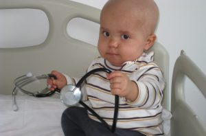 Primary immunodeficiencies (PIDs), also known as inborn errors of immunity (IEIs), are a group of diseases caused by the quantitative and/or functional alteration of different mechanisms involved in the immune response. The differences in their clinical and immunological manifestations, especially the type of infections they present with, are related to the molecular alteration in each case.
Patients with PID are susceptible to infections of varying severity which, if not treated appropriately, may be fatal or leave sequelae that worsen patients’ quality of life. In addition, more and more examples of these diseases are currently emerging in which there is selective susceptibility to certain microorganisms, which makes us increasingly alert to the existence of these conditions.
It is very important to bear in mind that primary immunodeficiencies are often associated with inflammation, immune dysregulation and neoplasms — especially of lymphoid tissue — at a much higher frequency than in the general population.
Primary immunodeficiencies occur in more than 1 in 2,000 live births, with great variability between the different conditions. Thus, while selective IgA deficiency is very common and affects 1 in 500 people in the Caucasian population, the group of severe combined immunodeficiencies (SCID) occurs in around 1 in 50,000 live births.
However, it is assumed to be a widely underdiagnosed group of disorders; some reviews indicate that only 10% of existing PIDs are diagnosed. It is estimated that PIDs, although classified individually as rare diseases, are collectively more common than well-known diseases such as cystic fibrosis and have a frequency very close to that of multiple sclerosis.
Despite this, there is still a great lack of awareness of them both among the general population and within the healthcare community.
Within primary immunodeficiencies, predominantly antibody deficiencies account for more than half of cases, and the next most frequent group is combined immunodeficiencies of T and B cells.
How are primary immunodeficiencies classified?
More than 550 primary immunodeficiencies have currently been described, of which the genetic defect is known in more than 450.
The current classification was drawn up by an international committee of experts, the International Union of Immunology Societies (IUIS), which at its latest biennial meeting in 2024 grouped PIDs into the following 10 groups:
- Combined immunodeficiencies of T and B cells
- Predominantly antibody deficiencies
- Combined immunodeficiencies with syndromic features
- Diseases of immune dysregulation
- Defects in phagocyte number and/or function
- Defects in innate immunity
- Complement deficiencies
- Autoinflammatory disorders
- Phenocopies of PID
- Bone marrow failure
What are the symptoms of primary immunodeficiencies?
Regarding the clinical suspicion index required to initiate the search for a primary immunodeficiency, there are warning signs that can guide us. Based on the classic warning signs proposed in 1993 by the Jeffrey Modell Foundation, new knowledge on IEIs has been incorporated, which, in addition to infections, includes autoimmune, inflammatory and neoplastic manifestations within the framework of the PIDCAP project.
| Warning signs defined by the PIDCAP programme in paediatric patients |
|---|
| 10 or more episodes of otitis media |
| 3 or more episodes of sinusitis or orbital cellulitis |
| 3 or more episodes of pneumonia |
| 2 or more months of antibiotic treatment per year |
| Growth and weight delay |
| Deep abscesses in organs |
| Lymph node abscess, recurrent skin abscesses (3 or more) or more superficial abscesses |
| Mucocutaneous candidiasis (oropharyngeal or cutaneous, excluding vaginal candidiasis) in children over 12 months of age: 2 or more episodes |
| 2 or more systemic infections, including sepsis |
| 5 or more hospital admissions |
| A single severe disease is sufficient to warrant investigation for primary immunodeficiencies (HSV meningitis, etc.) |
| Family history of primary immunodeficiencies |
| Consanguinity or other family history compatible with manifestations of primary immunodeficiencies (lymphomas, etc.) |
| Presence of cytopenia, whether or not it is specified as autoimmune |
| Autoimmune cytopenias |
| Systemic autoimmune diseases, excluding autoimmune cytopenias (coeliac disease, arthritis, etc.) |
| Endocrine disorders: hypothyroidism, hyperparathyroidism, diabetes, etc. (not described as autoimmune) |
| Haematological neoplasm |
| Solid-organ neoplasm (only those associated with primary immunodeficiencies in paediatric patients: thyroid) |
| Oral, dental or palatal abnormalities |
| Chronic diarrhoea or 10 or more recorded episodes of diarrhoea |
| 20 or more viral skin infections or chronic viral skin infection |
| Chronic eczema or other dermatological manifestations associated with primary immunodeficiencies |
| Recurrent fever |
| Inflammatory bowel disease in patients aged 2 years or older |
| Inflammatory bowel disease in children under 2 years of age |
| Presence of bronchiectasis without cystic fibrosis |
| Vaccine reaction |
| Presence of 2 or more items |
| Warning signs defined by the PIDCAP programme in adult patients |
|---|
| 8 or more episodes of acute otitis media |
| 8 or more episodes of sinusitis or chronic sinusitis |
| 3 or more episodes of pneumonia |
| Chronic diarrhoea and/or weight loss (10 or more episodes) |
| Deep abscesses in organs |
| Recurrent skin abscesses (more than 2) or superficial abscesses |
| Oropharyngeal or cutaneous candidiasis (excluding vaginal candidiasis) |
| Recurrent viral infections (colds, herpes, warts, condylomas, etc.): 25 or more episodes |
| Need for intravenous antibiotics to clear infections |
| 2 or more systemic infections, including sepsis |
| A single severe disease is sufficient to warrant investigation for primary immunodeficiencies |
| Infections caused by low-virulence bacteria belonging to the tuberculosis family |
| Family history compatible with primary immunodeficiencies |
| Consanguinity or other family history compatible with manifestations of primary immunodeficiencies, such as haematological neoplasms |
| Presence of cytopenia, whether or not it is specified as autoimmune |
| Presence of bronchiectasis without cystic fibrosis |
| Autoimmune cytopenias |
| Systemic and endocrine autoimmune diseases (coeliac disease, arthritis, SLE, thyroiditis, etc.) |
| Haematological neoplasm (excluding multiple myeloma, chronic myeloid leukaemia, Waldenström disease, etc.) |
| Solid-organ neoplasm associated with primary immunodeficiencies, specifically involving the skin, stomach or thyroid |
| Inflammatory bowel disease |
| Oral, dental and palatal abnormalities |
| Chronic eczema or other dermatological manifestations |
| Recurrent fever |
| Presence of 2 or more items |
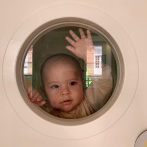 However, it is important to bear in mind that it is usually the combination of two or more of these signs and symptoms that leads to a real suspicion of a primary immunodeficiency. In addition, the weight of each variable is different. These warning signs exist for both children and adults.
How is a primary immunodeficiency diagnosed?
In Catalonia, since January 2017, universal screening for severe combined immunodeficiency has been carried out by determining T-cell receptor excision circles (TRECs) in the newborn heel-prick dried blood spot test.
For the remaining PIDs, the diagnostic process is based on clinical suspicion, complemented by the necessary laboratory tests. A complete clinical history must always be taken, collecting family data such as a history of PID or consanguinity, as well as the patient’s personal and pathological data, together with a thorough physical examination.
Analytical, immunological and genetic studies must be carried out in a rational and orderly manner in order to address the clinical suspicion and confirm the diagnosis.
In the case of genetic confirmation of a PID, it is essential to complete the study at family level in order to detect secondary cases and, where appropriate, offer suitable preconception counselling.
Since 2017, Catalonia has led a project using these warning signs — reviewed in the form of a computer algorithm — to guide primary care professionals in detecting possible primary immunodeficiencies earlier. This project, called PIDCAP, is currently in a phase of internationalisation and assessment for implementation in the Catalan health system.
At what age do the clinical manifestations of a primary immunodeficiency begin?
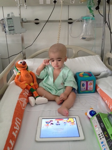 Although most primary immunodeficiencies present in childhood, they can manifest at any age and it is very important to also think of these entities in the adult patient. The age of onset of symptoms can also be used as a guide to one immune system defect or another.
By way of example, severe infections during the first months of life are indicative of primary combined immunodeficiency, however in predominantly humoral primary immunodeficiencies, infections usually begin after 5-6 months of life, at which time maternal immunoglobulins are no longer present.
Common variable immunodeficiency can present at any age, although as with IgA deficiency, the diagnosis cannot be confirmed until the patient has reached the age of 4 years. However, it should be kept in mind that these are only indicative data, as more and more “atypical” forms of certain PIDs are described that present milder and later than the classic forms.
How is a primary immunodeficiency diagnosed? Laboratory studies approach
As mentioned above, a large proportion of PIDs are diagnosed through a proper clinical history and physical examination, a full blood count and a determination of plasma immunoglobulins (Ig), tests that are available in most laboratories and in primary care. In any case, in recent years this diagnostic process has become more complex, with a predominant role for genetic studies.
Overall, in cases of suspected primary immunodeficiency, the initial study should include:
- A full blood count, with differential blood cell count, in which lymphopenia or neutropenia may be identified.
- Serum Ig levels (IgG, IgM and IgA): mainly to assess hypogammaglobulinaemia. Special situations involving differential diagnosis or targeted suspicion, in which IgE may be very useful, should also be considered.
A second diagnostic step would include:
- Determination of functional antibodies against protein antigens, such as tetanus, or polysaccharide antigens, such as pneumococcus or Salmonella typhi. These may be absent even when total Ig levels are previously found to be within the normal range.
- Basic lymphocyte immunophenotyping: CD3+ (T lymphocytes), CD3+CD4+, CD3+CD8+, CD4/CD8 ratio, CD19 (B lymphocytes) and CD56+ (NK).
- Advanced lymphocyte immunophenotyping with T- and B-lymphocyte subpopulations.
- Lymphocyte proliferative function. This assesses the ability of lymphocytes to react in vitro to different stimuli in order to draw a parallel with what could occur in vivo. It is used to detect cases of combined immunodeficiencies, although its role in the diagnosis of PIDs has diminished in recent years.
- Genetic studies (Sanger sequencing, PID panels, exome, genome, array-CGH, etc.).
- Functional studies to validate genetic results when they are inconclusive.
- In cases of suspected specific PIDs, tests such as the granulocyte oxidative burst test by flow cytometry — to rule out chronic granulomatous disease — or CH50 and AP50 determination to assess the complement pathway, among others, may be considered.
It is important to assess the results according to the reference values for each age, as there are significant differences which, if not taken into account, may lead to overlooking the diagnostic suspicion of primary immunodeficiency.
Guidance is always important, depending on the diagnostic suspicion, to avoid unnecessary tests that waste time and resources. However, when the diagnosis is uncertain and suspicion is high, additional tests such as functional or molecular studies are necessary and should be performed at reference centres.
What is the treatment for primary immunodeficiencies?

×
Advances in knowledge regarding aetiology, clinical and therapeutic aspects have profoundly improved the prognosis of these patients in recent years. The treatment of patients with primary immunodeficiency is divided into the so-called support treatment and curative treatments as described below.
Support treatment
- Immunoglobulin (Igs) replacement therapy:
Igs are plasma preparations drawn from a pool of thousands of donors. They contain neutralising antibodies against numerous viral and bacterial pathogens. They undergo sterilisation processes that eliminate other proteins and live viruses such as HBV, HIV, HCV.
It is the treatment of choice for patients with significant alterations in humoral immunity such as common variable immunodeficiency (CVID), X-linked agammaglobulinemia (XLA), and antibody production defects, as well as combined immunodeficiencies and many other immunodeficiency-associated syndromes.
They can be administered intravenously or subcutaneously and reduce the incidence and severity of infections, thereby reducing morbidity and mortality in these patients.
This requires maintaining serum IgG trough concentrations above 600-800 mg/dl before further administration, although both the dose and the trough concentrations to be obtained must be individualised for each patient according to their infection rate.
- Antimicrobial treatment:
Antimicrobial therapy should be initiated early in patients with infectious manifestations. Adequate cultures are essential because of the possibility of opportunistic pathogens that are common in patients with primary immunodeficiencies.
Prophylactic antibiotics, not routinely recommended, are sometimes indicated to protect the patient from pathogens to which he or she may be susceptible. An example of this is the treatment with trimethoprim-sulfamethoxazole in patients with hyper IgM syndrome and in those with T-cell defects following haematopoietic precursor transplantation for protection against Pneumocystis jirovecii pneumonia.
Primary antimicrobial prophylaxis is essential in phagocytic defects and should be considered for complement defects and in hyper IgE syndrome. In certain PIDs with a high susceptibility to bacterial, fungal or viral infections, prolonged antimicrobial prophylaxis is necessary and should be assessed on an individual basis.
- Other therapies:
Increasingly, immunomodulatory or immunosuppressive treatments are used to treat the inflammatory or autoimmune manifestations that these patients may present. If the genetic defect is known, this may allow the use of targeted treatments aimed at optimising the benefit-risk ratio (leniolisib in APDS types I and II, abatacept in CTLA-4 or LRBA, sirolimus in IPEX, etc.).
Enzyme replacement has been used in ADA deficiency, the first human enzyme deficiency to be treated with replacement therapy.
In addition, interferon gamma has been used as a treatment in chronic granulomatous disease and immunisations against encapsulated microorganisms are recommended in certain complement deficiencies. Plasma concentrate derivative of recombinant C1 esterase inhibitor is available for the treatment of hereditary angioedema.
Curative treatments
- Haematopoietic precursor transplantation (bone marrow transplantation):
Haematopoietic precursor transplantation (bone marrow transplantation) is, together with gene therapy, the only curative option for many primary immunodeficiencies.
It is the treatment of choice for some immunodeficiencies such as severe combined immunodeficiency (SCID), Wiskott-Aldrich syndrome, chronic granulomatous disease, among others.
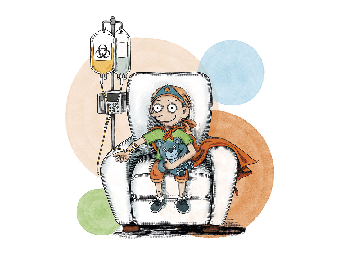
In patients with severe combined immunodeficiency (SCID) who receive a transplant from an HLA-identical donor, survival rates reach 90%; this is similar to when the transplant is from a non-familial but matched donor and performed very early. Early diagnosis and the search for optimal donors, together with conditioning, where appropriate, achieves more favourable outcomes.
- Gene therapy:
Gene therapy is becoming a reality for many primary immunodeficiencies such as Wiskott-Aldrich syndrome, ADA deficiency, some forms of SCID, some forms of chronic granulomatous disease and others.
Although several PIDs have been successfully treated in early trials, the outcome of this therapy is mixed and, unfortunately, several patients have subsequently developed myelodysplastic syndrome associated with the activation of proto-oncogenes by the vector.
With the use of new vectors in the last decade, multiple successful gene therapies have been performed without these undesirable effects and are now a viable option for a variety of PIDs.
What are the chances of children with a severe combined immunodeficiency being cured?
Without treatment, severe combined immunodeficiency (SCID) often leads to severe infection and death in children before their first birthday.
If bone marrow transplantation is performed in the first few months of life from a compatible donor, it can offer a long-term survival rate of over 90%, especially in the absence of pre-transplant infections. This option is now a reality in our setting thanks to newborn screening for these conditions and improvements in the transplant procedure.
Links of interest concerning medical issues relating to primary immunodeficiencies
Information on severe combined immunodeficiency. National Center for Advancing Translational Sciences
We recommend visiting the following websites of specialised foundations:
If you have PID or you are a family member, you can also access the different family associations in Spain:
Links of interest on other topics related to primary immunodeficiencies
All these organisations are external to the Josep Carreras Leukaemia Foundation.
ANDALUCÍA
ARAGÓN
ASTURIAS
CASTILLA LA MANCHA
CASTILLA LEÓN
CATALUÑA
VALENCIAN COMMUNITY
EXTREMADURA
GALICIA
BALEARIC ISLANDS
CANARY ISLANDS
LA RIOJA
MADRID
- AAA (asociación de adolescentes y Adultos Jóvenes con Cáncer)
- ASION
- FUNDACIÓN CAICO
- FUNDACIÓN ALADINA
- FUNDACIÓN UNOENTRECIENMIL
MURCIA
NAVARRA
BASQUE COUNTRY
Support and assistance
We also invite you to follow us through our main social media (Facebook, Twitter and Instagram) where we often share testimonies of overcoming this disease.
If you live in Spain, you can also contact us by sending an e-mail to imparables@fcarreras.es so that we can help you get in touch with other people who have overcome this disease.